|
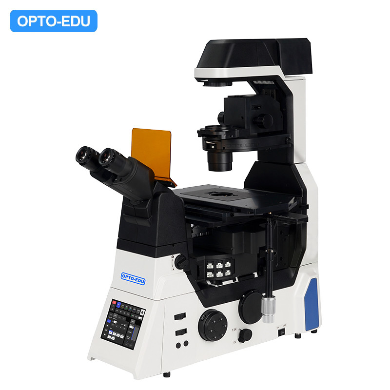
A16.1099-A Auto Motorized Scientific Research Grade Inverted Fluorescent Microscope
It could realize high-speed, full-automatic component linkage, compatible with adaptive focusing systems (AFS), especially suitable for high-level living cell research. double layer optical path provides more scalability possibilities, providing perfect flexible and open inverted microscopic imaging platform for advanced microteaching such as living cell, confocal, super resolution. |
|
A16.1099-M Manual Coded Scientific Research Grade Inverted Fluorescent Microscope
Scientific research grade inverted microscope for advanced life science research, integrating bright field, phase contrast, Hoffman, DIC, fluorescence etc. observation methods. Intelligent coding components record interactive information and workflow, Z-axis motorized focusing can simplify focusing steps, reduce visual fatigue and improve work efficiency in daily research, teaching and pathological diagnosis. |
|
▶ Incomparable 25mm Field of View
With the research trend towards large-scale, high-flux and intelligent solutions, demand for faster data acquisition and higher throughput is increasing day by day. No matter it is bright field imaging or fluorescence imaging,A16.1099 large-area illuminator and large-field fluorescence attachment provides uniform and bright cell imaging. Large size sensors and imaging interfaces that truly maximize performance of large format detector and provide perfect imaging platform for the future as camera technology continues to evolve rapidly. |
|
▶ Large field illumination module |
|
|
Bright and large area transmission illumination Transmission illumination module adopts high power LED illumination to guarantee full field of view brightness and uniform illumination, ensure get clear, consistent results from high magnification DIC applications, provide stable light source for seamless image stitching. |
Large aperture reflective fluorescent illuminator A compact optical fiber illumination device for FOV25mm fluorescence imaging is designed,, equipped with high power LED light box, provides wide spectrum high transmittance lighting including ultraviolet light, meanwhile compatible with large aperture fluorescent filters, provide fluorescence images of FOV25mm with high signal-to-noise ratio. |
|
▶ Large aperture observation optical system Equipped with large diameter tube lens to expand light flow, cooperate with large target surface CMOS sensor, both bright field and fluorescence imaging could reach FOV25mm field of view. |
▶ FOV25 imaging objective The objective with superior image flatness guarantees high quality images. Maximize potential of FOV25 objectives significantly speeds up data collection. |
|
▶ Camera for large volume data acquisition NEXCAM-MAX2400 High sensitivity monochrome camera, full frame sensitive Chip, target surface 36.0x23.9mm, 24 millions pixel CMOS Image sensor, maximum acquisition speed could reach 114fps, realize digital imaging of the largest field of vision. |
|
|
▶ Ideal Living Cells Microscopic Experiments Platform
Even small changes of temperature and vibration in imaging environment can greatly affect stability of focus, adaptive anti-focus shift system (AFS) eliminates focus drift through static and dynamic measurements, high brightness LED fluorescent light source and live cell culture system enables A16.1099 realize multi-day living cell time-lapse observation, making it an ideal imaging tool for more demanding focused experiments. |
|
▶ High Specification LED Fluorescent Illumination System LED 4 enables up to 4 channels LED illumination, highly matching fluorescent dye commonly used in market, excitation energy concentrated, high brightness, meet the daily experimental fluorescence imaging need. long life, do not need to change bulb. Compared to traditional mercury lamps, it reduce photobleaching and phototoxicity, which are very friendly to live cell samples .It is an ideal microscope light source which is sustainable, energy saving and low carbon, environmental protection. |
|
▶ Intuitive and ergonomic control operation
A16.1099 integrates a large number of advanced human-computer interaction technologies, which enables fine control of imaging. Intuitive and easy microscope operation interface can make the complex operation procedures become very simple, so that researchers can work more efficiently and comfortably, which helps to reduce researchers’ fatigue while the system minimizes damage to living cells.. |
|
▶ Interactive operation A16.1099 was creatively designed front panel as a touch screen, which makes human-computer interaction more convenient, powerful and expandable. Left and right sides reserved knobs and buttons, even in a dark lab, it is easy to control, which allows researchers to focus more on the experiment itself, rather than complex microscope operation. |
|
Front 5.6-inch touch screen Touch screen control movement of electric parts such as objective, double layer/single layer fluorescent turnable, conden5.6-inchsity, motorized platform speed, motorized Z-axis speed, main body splitting port, ESC escape, FN key, objective focus. Display real-time, display objective magnification, transmitted illumination brightness, fluorescence band, output port, XYZ position and speed and other component status. |
|
|
▶ High speed electric control Operation and conversion speed of objectives, filters, the XY stage and the observation module are greatly improved, enabling an easy operating environment for researchers to concentrate on routine observations and images. The joystick that can control the stage freely allows the microscope working as your eyes and hands, making it easy to use. |
|
Joystick with built-in touch panel The joystick comes with touch screen, which can set the state of main body, motorized platform control by multi-level and also motorized focus, built-in function key and AFS focus stabilization control. |
|
|
A16.1099 high quality infinity optical system, equipped with bright field, fluorescence, phase contrast, DIC, Hoffmann and other complex observation methods, no matter which observation method, you can get bright high signal-to-noise ratio of the original image, with its excellent optical performance and reliable reliability are highly valued by researchers |
|
|
Phase contrast The condenser has built-in phase contrast ring, which can be converted from bright field observation to phase contrast observation by turning the condenser turntable. Semi apochromatic phase contrast objective provides clear imaging with high contrast. |
Fluorescence Using the newly designed filter group, cutoff depth of single channel fluorescence filter reaches OD6, and the image with high signal-to-noise ratio can be obtained, which can be used for single-molecule and single-cell experiments. illumination module spectrum design covers a wide wavelength range, and six-hole filter module turntable meets needs of multi-color fluorescence. High power LED fiber optic light source is bright and safe. |
|
DIC(Differential interference) High quality large field of view differential interference optics system covers all magnifications, providing uniformly clear and detailed images with high resolution and contrast for each sample. |
|
|
|
Hoffmann contrast High contrast ratio phase contrast imaging technique for unstained, transparent samples such as egg cells. Hoffmann phase contrast components provide pseudo-three-dimensional image with the appearance of shadow casting. The contrast direction of each sample can be easily adjusted. |
| Research Level Inverted Fluorescent Microscope A16.1099 | -M | -A | Cata. No. | |
| Main Frame | Manual Frame | ● | - | |
| --5.7" Touch Screen, Show All Status, Control Only Light Split | ||||
| --Motorized Z Axis Control | ||||
| --Coded Brightness Adjust Knob | ||||
| --Coded Manual Intermediate Magnification 1x/1.5x Switch Button | ||||
| --Coded Manual Bertrand Lens Switch | ||||
| Motorized Frame | - | ● | ||
| --5.7" Touch Screen, Show All Status, Control All Parts | ||||
| --Motorized XYZ Axis Control | ||||
| --Coded Brightness Adjust Knob | ||||
| --Coded Manual Intermediate Magnification 1x/1.5x Switch Button | ||||
| --Coded Manual Bertrand Lens Switch | ||||
| --F1 Button For Customized Function | ||||
| --Motorized Nosepiece Objective Switch Button x2 | ||||
| --Motorized Condenser Button | ||||
| --Motorized Fluorescent Filters Switch Wheel x2 | ||||
| Eyepiece | WF10x/22mm Eyepiece, Diopter Adjustable +/-5°, High Eyepoint | ●● | ●● | A51.1006-1022A |
| Head | 10-40° Tilt Adjustable Binocular Head, IPD 47-78mm, Eyepiece Tube Dia. 30mm | ● | ● | A51.1099-B |
| Mag. Switch | Coded Manual Intermediate Magnification Switch Button 1x/1.5x On Right Side | ● | ● | |
| Nosepiece | Manual 6 Holes Nosepiece, Coded, With DIC Slot, M25×0.75 | ● | ||
| Motorized 6 Holes Nosepiece, With DIC Slot, M25×0.75 | ● | |||
| Objective | Infinity Plan Semi-APO Phase Contrast 10x, N.A.0.3, W.D.15.7mm, Cover Glass 1.2mm | ● | ● | A5C.1099-10 |
| Infinity Plan Semi-APO Phase Contrast 20x, N.A.0.45, W.D.8mm, Cover Glass 1.2mm | ● | ● | A5C.1099-20 | |
| Infinity Plan Semi-APO Phase Contrast 40x, N.A.0.6, W.D.3.6mm, Cover Glass 1.2mm | ● | ● | A5C.1099-40 | |
| Working Stage | Manual Mechanical Stage, 3 Layers, XY Moving 130×85mm, Size 340x230mm, Flexiable Long Handel, Can Fit Different Holder On Top | ● | - | |
| Petri Dish Holder Size 160(X)×110(Y)mm | ● | - | ||
| Petri Dish Holder Size 81(X)×55.5(Y)mm | ● | - | ||
| Slide Holder Size 160(X)×110(Y)mm | ● | - | ||
| 96 Holes Plate Holder Size 160(X)×110(Y)mm | ● | - | ||
| Motorized Working Stage, Grating Type, XY Moving 130x100mm, Size 445x300mm, Max Speed 20mm/s, Moving Precision 0.1μm, Repeatability 0.5μm | - | ● | ||
| Slide & Petri Dish Holder For Dia. 35-65mm | - | ● | ||
| 4~1396 Holes Plate Holder | - | ● | ||
| Focusing | Motorized Z Axis, Grating Type, Moving Range Up 8.5mm, Down 1.5mm, Min Step 0.02um, Repeatability 0.1um, 3 Level Focusing Knob: 2um/40um/200um Per Circle | ● | ● | |
| Transmit Light | Kohler Illumination, With Field/Iris Diaphragm, 0~25° Titl Adjustable Arm | ● | ● | |
| Electrical Optical Brake Optional | ||||
| 3W S-LED Lamp House | ● | ● | ||
| Condenser | Manual 7 Holes Nosepiece Turret, Coded, 4 Holes For Phase Contrast, Hoffman, ND Filter, 3 Holes For DIC, ND Filters, Up/Down Range 66mm | ● | ||
| Motorized 7 Holes Nosepiece Turret, 4 Holes For Phase Contrast, Hoffman, ND Filter, 3 Holes For DIC, ND Filters, Up/Down Range 66mm | ● | |||
| Long Working Distance Condenser NA=0.52,WD=30mm | ● | ● | ||
| Phase Contrast | 10x-20x Phase Contrast Module | ● | ● | |
| 40x Phase Contrast Module | ● | ● | ||
| Coded Manual Bertrand Lens Switch In/Out Light Path, Focus Adjustable | ● | ● | ||
| DIC | DIC Prism Slide (Into Nosepiece DIC Slot), DIC Polarizer Slide, DIC Analyzer Slide | o | o | A5C.1099 |
| Reflect Light | Epi Fluorescent LED Light Source, Optical Fiber Adapter, Iris Diaphragm, 2 Holes Filter Insert Plate | ● | ● | |
| Round Diaphragm | ● | ● | ||
| Support 4 Fluorescent LED Light Source: 385, 470, 555, 630 | ● | ● | ||
| Light Source Controller Box | ● | ● | ||
| Fluorescent Turret | Manual 6 Holes Fluorescent Turret Disc, Manual Shutter, With Cable | ● | ||
| Motrozied 6 Holes Fluorescent Turret Disc, Motorized Shutter, With Cable | ● | |||
| FITC Filter, BP460-495, DM505, BA510-550 | ● | ● | ||
| TRTC Filter, BP528-553, DM565, BA578-633 | ● | ● | ||
| DAPI Filter, BP360-390, DM415, BA435-485 | ● | ● | ||
| Double Layer Fluorescent Turret Set, Including | o | o | A5F.1099 | |
| Z Axis Heighten Base, Fluorescent Turnable Heighten Seat, Stage Heighten Seat | ||||
| Middle Part | For Light Split Status Detection: 100/0, 0/100 | ● | ● | |
| Photo Adapter | Motorized Light Split Switch Control By Front 5.7" Touch Screen: | ● | ● | |
| --Eyepiece 100%, | ||||
| --Left Photo Port 100%, | ||||
| --Right Photo Port 100%, | ||||
| --Eyepiece 20% + Right Photo Port 80% | ||||
| Left Photo Port, With Built-in 1x C-Mount | ● | ● | A55.1099-L1.0 | |
| 0.7x C-Mount For Left Photo Port | o | o | A55.1099-L0.7 | |
| 0.5x C-Mount For Left Photo Port | o | o | A55.1099-L0.5 | |
| 1x C-Mount For Right Photo Port, | - | ● | A55.1099-R1.0 | |
| Confocal | Left Side Confocal Port With 1x Lens | ● | ● | |
| Joystick | Joystick For XYZ Motorized Control, Touch Screen Show Objectives, Fluorscent Filters, Customized Shortcut Button | ● | ||
| Tool Kit | Full Set Tool Kit For Installation | ● | ● | |
| Dust Cover | Dust Cover | ● | ● | |
| Cable | USB2.0 Cable 1.5m, Motorized Control Data Cable | ● | ● | |
| Software | Support Image & Video Capture, Control Microscope XYZ Stage, Objectives Switch, Image & Video Capture, Large Image Stitching, Extended Depth of Field (EDF), | ● | ● | |
| Not Support Slide Scan Function | ||||
|
Adaptive Anti-Focus Shift System (AFS) Automatically correct focus drift caused by temperature changes and mechanical vibrations. |
Stage lifting position device Stage can be raised to allow installation of a second fluorescent illuminator and filter module turntable. |
Fluorescent filter module turntable Both manual coding model and electric intelligent model are available, compatible with FOV25 wide field of view. |
|
Electric platform High-speed electric platform with grating ruler for accurate positioning and high repeatability. |
Manual platform Long working stroke, can be a complete observation of the entire hole plate, a variety of platform holders are available. |
Live cell culture system Provide best conditions for the survival of living cells include precise temperature, humidity and gases. |
|
Fluorescent LED light source With high matching with fluorescent dyes commonly used in the market, which is ready to use, long life and more environmentally friendly. |
Condenser turnable Both manual model and electric model are available, it can automatically switch between seven positions to adapt to different imaging modes |
|
|
▶ A65.1099 Super Resolution Microscope
It can be used as imaging platform of A65.1099 laser confocal microscope to obtain low noise, high contrast and high quality confocal images. Equipped with SIM super resolution system, A65.1099 enables live cell imaging with twice the resolution of traditional optical microscopes. |
|
▶ A64.1099 Laser Confocal Microscope
Integrating various microscopical techniques such as brightfield, fluorescence, differential interference contrast, and phase contrast, users can freely opt for single-layer or dual-layer optical path configurations based on their specific experimental needs to achieve optimal imaging results. The Adaptive Focus Shift System (AFS) ensures precise focal plane positioning during continuous observations, thereby enabling stable, continuous, and clear recordings of cellular dynamic behaviors. |
Our products are sold all over the world, you can rest assured.