|
▶ High Speed Hardware Control
It empowers users with unprecedented convenience in operation, effortlessly digitizing management and enabling precise control over multiple electric components within the microscope, such as objective lens switching, focusing, condenser lens changing, and fluorescence module transitions. |
|
▶ Multi Dimensional Imaging And Display
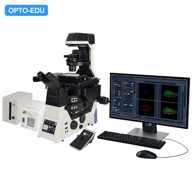
It is capable of memorizing custom observation modes and supports the combined use of X, Y, Z, λ, and T scanning functions. Equipped with a variety of flexible shooting modes, including multi-channel fluorescence imaging, time-lapse scanning, multi-position acquisition, Z-axis stacking, and panoramic stitching. These five modes can be freely combined according to the user's actual needs, adapting to a wide range of complex and diverse experimental application scenarios. |
|
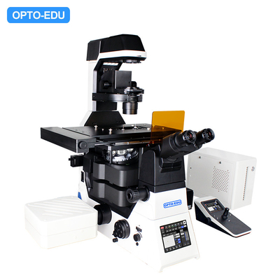
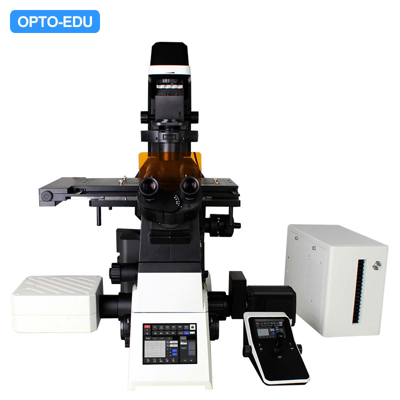
▶ High Performance Confocal Microscopy Platform The A16.1099 offers a powerful and flexible imaging solution, establishing a robust and highly expandable foundation for microscopic imaging within the A64.1099 system. With its 25mm field of view design, it provides ideal observation conditions for large sample and high-throughput experimental research. Integrating various microscopical techniques such as brightfield, fluorescence, differential interference contrast, and phase contrast, users can freely opt for single-layer or dual-layer optical path configurations based on their specific experimental needs to achieve optimal imaging results. The Adaptive Focus Shift System (AFS) ensures precise focal plane positioning during continuous observations, thereby enabling stable, continuous, and clear recordings of cellular dynamic behaviors. |
|
▶ High Speed Electric Motor Control The operation and switching speed of objectives, filter blocks, XY stage, and observation modules have been significantly enhanced, creating an effortless operating environment that enables researchers to focus on daily observations and image capture. A joystick for intuitive manipulation of the stage allows the microscope to become an extension of your eyes and hands, making it user-friendly and natural to operate. |
|
▶ Live Cell Culture System Specifically designed for precise live cell imaging, this system accurately regulates the microscope platform temperature, ensuring constant temperature, humidity, and CO2 concentration within the culture dish, thereby providing an ideal cultivation environment for long-term experiments. |
|
|
▶ AFS Ensures Stable And Reliable Imaging Performance. The A16.1099 employs an independent focusing design, minimizing the impact of other mechanical components on the Z-axis. It features a newly designed Adaptive Focus System (AFS), which intelligently eliminates focus drift. Whether paired with high-magnification objectives has large numerical apertures or utilized in conjunction with advanced imaging techniques such as super-resolution, confocal, or TIRF (Total Internal Reflection Fluorescence), the system consistently delivers crisp, sharp images. This design ensures the highest level of imaging stability and precision across a broad range of demanding applications in modern microscopy.
|
|
|
▶ High Power LED Fluorescence Illuminator The LED 4 system enables up to 4-channel LED illumination, offering high compatibility with commonly used fluorescent dyes in the market. It features concentrated excitation energy and high brightness, fulfilling the fluorescence imaging requirements of routine experiments. With instant-on capability, long service life, and no need for bulb replacement, it outperforms traditional mercury arc lamps in terms of reducing photobleaching and phototoxicity, making it particularly friendly to live cell samples. It is a sustainable, energy-efficient, and environmentally friendly microscope light source, ideal for low-carbon laboratory practices. |
|
|
▶ Smart Interactive Operation The A16.1099 innovatively incorporates a touch screen into its front panel, significantly enhancing user interface convenience and expandability of functions. It retain the traditional microscope knobs and buttons on both sides ensures intuitive control even in dark laboratory environments, allowing researchers to concentrate on the core of their experiments without being hindered by complicated operations. This design promotes an efficient and seamless microscopy observation experience. |
|
A64.1099 is equipped with a 5.6-inch touch screen display on the front panel. Control of components such as objectives, dual-layer/single layer fluorescence filter wheels, the condenser, light intensity, electric stage speed, electric Z-axis speed, host spectrometer ports, ESC exit, FN keys, and objective parfocality is achieved through the touch interface. It also provides real-time display of various statuses including objective magnification, transmitted illumination brightness, fluorescence wavelengths, output ports, XYZ positions, and movement speeds. |
|
|
▶ Large Aperture Observation Optical System Equipped with a large-aperture objective lens, it significantly increases light transmission, coupled with a spacious CMOS sensor, effortlessly enabling brightfield and fluorescence imaging across a vast field of view up to FOV25mm. This broader perspective captures more details, empowering you to comprehensively explore the microscopic world and have complete control over your scientific research endeavors. |
|
|
▶ Large Aperture Reflective Fluorescence Illuminator Specifically designed for a large field of view (FOV) of 25mm, this fluorescence imaging illumination apparatus features a high-power LED light box, delivering broadband, high-transmission illumination encompassing the ultraviolet spectrum. It is also compatible with large-aperture fluorescent filters, ensuring high signal-to-noise ratio fluorescent images for detailed and accurate observations. |
|
| A64.1099 Laser Confocal Microscope | Cata. No. | ||
| Main Frame | Motorized Frame | ● | |
| --5.7" Touch Screen, Show All Status, Control All Parts | |||
| --Motorized Z Axis Control | |||
| --Coded Brightness Adjust Knob | |||
| --Coded Manual Intermediate Magnification 1x/1.5x Switch Button | |||
| --Coded Manual Bertrand Lens Switch | |||
| --F1 Button For Customized Function | |||
| --Motorized Nosepiece Objective Switch Button x2 | |||
| --Motorized Condenser Button | |||
| --Motorized Fluorescent Filters Switch Wheel x2 | |||
| Eyepiece | WF10x/22mm Eyepiece, Diopter Adjustable +/-5°, High Eyepoint | ●● | A51.1006-1022A |
| Head | 10-40° Tilt Adjustable Binocular Head, IPD 47-78mm, Eyepiece Tube Dia. 30mm | ● | A51.1099-B |
| Mag. Switch | Coded Manual Intermediate Magnification Switch Button 1x/1.5x On Right Side | ● | |
| Nosepiece | Motorized 6 Holes Nosepiece, With DIC Slot, M25×0.75 | ● | A54.1099-M |
| Motorized 6 Holes Nosepiece, With Adaptive Anti-Focus Shift System (AFS) | o | A54.1099-MAFS | |
| Objective | NIS60 Infinity Plan APO Achromatic Objective 4x, N.A.0.16, W.D.17mm | o | A52.1099-4 |
| NIS60 Infinity Plan APO Achromatic Objective 10x, N.A.0.45, W.D.4mm | ● | A52.1099-10 | |
| NIS60 Infinity Plan APO Achromatic Objective 20x, N.A.0.75, W.D.1.1mm | ● | A52.1099-20 | |
| NIS60 Infinity Plan APO Achromatic Objective 40x, N.A.0.95, W.D.019-0.21mm | ● | A52.1099-40 | |
| NIS60 Infinity Plan APO Achromatic Objective610x (Oil), N.A.1.42, W.D.0.25mm | ● | A52.1099-100 | |
| NIS60 Infinity Plan APO Achromatic Objective 100x(Oil), N.A.1.45, W.D.0.13mm | o | A52.1099-100 | |
| NIS60 Infinity Plan APO Achromatic Objective 100x(Oil), N.A.1.49, W.D.0.09-0.16mm, Cover Glass 0.13~0.19(23C°), 0.14~0.20(37°C) | o | A52.1099-100A | |
| Motorized Working Stage, Grating Type, XY Moving 130x100mm, Size 445x300mm, Max Speed 20mm/s, Moving Precision 0.1μm, Repeatability 0.5μm | ● | ||
| Slide & Petri Dish Holder For Dia. 35-65mm | ● | ||
| 4~1396 Holes Plate Holder | ● | ||
| Holder For 96 Holes Petri Dish | o | A54.1099-96 | |
| Holer For For Olympus 6 Holes Petri Dish | o | A54.1099-6 | |
| Platform | Vibration Isolation Platform, 1000x1000mm | o | A54.1080 |
| Focusing | Motorized Z Axis, Grating Type, Moving Range Up 8.5mm, Down 1.5mm, Min Step 0.02um, Repeatability 0.1um, 3 Level Focusing Knob: 2um/40um/200um Per Circle | ● | |
| Transmit Light | Kohler Illumination, With Field/Iris Diaphragm, 0~25° Titl Adjustable Arm | ● | |
| Electrical Optical Brake Optional | |||
| 3W S-LED Lamp House | ● | ||
| Condenser | Motorized 7 Holes Nosepiece Turret, 4 Holes For Phase Contrast, Hoffman, ND Filter, 3 Holes For DIC, ND Filters, Up/Down Range 66mm | ● | |
| Long Working Distance Condenser NA=0.52,WD=30mm | ● | ||
| Phase Contrast | 10x-20x Phase Contrast Module | ● | |
| 40x Phase Contrast Module | ● | ||
| Coded Manual Bertrand Lens Switch In/Out Light Path, Focus Adjustable | ● | ||
| DIC & Polarizing | DIC Set, Including Manual Splitter, With Optical Fiber, View Field 18mm, With DIC Prism Slide (Into Nosepiece DIC Slot), DIC Polarizer Slide, DIC Analyzer Slide | ● | A5C.1099 |
| 10x DIC Nocaridan Prism Slide, Insert In Nosepiece DIC Slot | ● | ||
| 20x DIC Nocaridan Prism Slide, Insert In Nosepiece DIC Slot | ● | ||
| 40x DIC Nocaridan Prism Slide, Insert In Nosepiece DIC Slot | ● | ||
| 60x DIC Nocaridan Prism Slide, Insert In Nosepiece DIC Slot | ● | ||
| 100x DIC Nocaridan Prism Slide, Insert In Nosepiece DIC Slot | o | ||
| 10x DIC Walker Prism, In Condenser Turret | ● | ||
| 20x/40x/60x DIC Walker Prism, In Condenser Turret | ● | ||
| 100x DIC Walker Prism, In Condenser Turret | o | ||
| Reflect Light | Epi Fluorescent LED Light Source, Optical Fiber Adapter, Iris Diaphragm, 2 Holes Filter Insert Plate | ● | |
| Round Diaphragm | ● | ||
| Support 4 Fluorescent LED Light Source: 385, 470, 555, 630 | ● | ||
| Light Source Controller Box | ● | ||
| Fluorescent Turret | Motrozied 6 Holes Fluorescent Turret Disc, Motorized Shutter, With Cable | ● | |
| FITC Filter, BP460-495, DM505, BA510-550 | ● | ||
| TRTC Filter, BP528-553, DM565, BA578-633 | ● | ||
| DAPI Filter, BP360-390, DM415, BA435-485 | ● | ||
| Double Layer Fluorescent Turret Set, Including | o | A5F.1099 | |
| Z Axis Heighten Base, Fluorescent Turnable Heighten Seat, Stage Heighten Seat | |||
| Middle Part | For Light Split Status Detection: 100/0, 0/100 | ● | |
| Photo Adapter | Motorized Light Split Switch Control By Front 5.7" Touch Screen: | ● | |
| --Eyepiece 100%, | |||
| --Left Photo Port 100%, | |||
| --Right Photo Port 100%, | |||
| --Eyepiece 20% + Right Photo Port 80% | |||
| Left Photo Port, With Built-in 1x C-Mount | ● | A55.1099-L1.0 | |
| 0.7x C-Mount For Left Photo Port | o | A55.1099-L0.7 | |
| 0.5x C-Mount For Left Photo Port | o | A55.1099-L0.5 | |
| 1x C-Mount For Right Photo Port, | ● | A55.1099-R1.0 | |
| Laser Confocal | Left Side Confocal Port With 1x Lens | ● | |
| Galvanometer Scanning Mirror, Motorized Pinhole Stepless Adjustment. | ● | ||
| 4 Laser Source With AOTF, Max Laser Power 50mW, | ● | ||
| 405nm, 488nm, 561nm, 640nm | |||
| 4 PMT Detection, Captuer Set, Control Set, | ● | ||
| EX 405,488,561,640 | |||
| Channel 1 EM:440-470 | |||
| Channel 2 EM:510-540 | |||
| Channel 3 EM:575-605 | |||
| Channel 4 EM:679-727 | |||
| Scan Head Max Pixel 8192x8192 (8K x8K) | ● | ||
| Scan Mode Support Combine Use of X, Y, Z, λ, T Scanning Functions | |||
| Confocal Field of View: Square Inscribed In Dia. 25mm Circle | |||
| Image Bit Depth: 16bits | |||
| Computer | i7-11700/16GB*2 DDR4/1TB SSD/RTX A2000 6G/USB PCIE Card, 4x USB3.0, WINDOWS 10 Professional, 3NBD/460W/ CD Driver, 4K Monitor | ● | |
| Camera | 1.1" CMOS, Work With 1x C-Mount, FOV 18mm, 4K, FPS 23.4, Max FPS 46.3 | ● | |
| 24M Digital Camera, 2.7" CMOS, Work With 1.5x C-Mount (M52), 6K High Resolution, 15.3FPS@20M, Max FPS 114 | o | A55.1099-24M | |
| Joystick | Joystick For XYZ Motorized Control, Touch Screen Show Objectives, Fluorscent Filters, Customized Shortcut Button | ● | |
| Tool Kit | Full Set Tool Kit For Installation | ● | |
| Dust Cover | Dust Cover | ● | |
| Cable | USB2.0 Cable 1.5m, Motorized Control Data Cable | ● | |
| Software | Support Image & Video Capture, Control Microscope XYZ Stage, Objectives Switch, Image & Video Capture, Large Image Stitching, Extended Depth of Field (EDF), NOT Support Slide Scan Function | ● | |
| Multimodal | Ultra-Wide Field of View, Super-Resolution, Ultra-High Speed |
| >Brightfield, Phase Contrast, Fluorescence, DIC | >Field of View reaching up to 66μm x 66μm at 100X Magnification |
| >Laser Confocal Microscopy | >Lateral Resolution (2D-SIM) improved 2-fold, with a maximum achievable resolution of 86nm; Axial Super-Resolution (3D-SIM) advanced to 270nm. |
| >2D-SIM/3D-SIM/TIRF-SIM | >Real-time super-resolution imaging at video rate, delivering "what you see is what you get" experience. |
Our products are sold all over the world, you can rest assured.